What is Digital Dentistry?
by Paphos Smile Art Studio Paphos Dental Clinic
Digital dentistry encompasses any digital or computer-based technology that your dental professional may use to examine, diagnose, and treat the health of your mouth.
 - Copy.jpg) |
Types of Digital Dentistry Used Today
by Paphos Smile Art Studio Paphos Dental Clinic
Digital dentistry is transforming just about every aspect of professional oral care from the moment you check in to your appointment to when your dental professionals assess your oral health, to the diagnosis and treatment of any conditions or diseases you may have, to how your dental professional follows-up and interacts with you between appointments. Some digital dentistry tools that you may come across in your appointments include:
Intraoral cameras
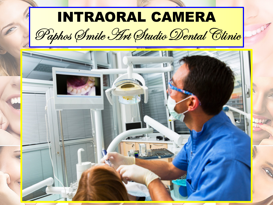
Small cameras are fast-replacing the tiny round mirrors dental professionals have historically used to examine the inside of your mouth. One of the biggest benefits of these cameras is magnification. When they can make your tooth about your head's size on a flat screen, they can better identify any potential issues with your oral health that need to be addressed. Another benefit is that they can share what they see with you, allowing you to better understand and improve your oral hygiene. Images can also be shared with lab technicians to match crowns and bridges to the shade of your actual teeth.
Digital radiography
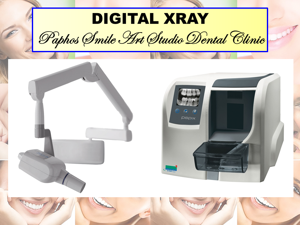
X-rays have been used in dental offices for a very long time, but the traditional x-ray process required film processing, which took time, was expensive, and the prints needed to be filed away in cabinets and physically delivered to other offices and specialists if necessary. Digital radiography is faster. The images are immediately available to see on a computer screen. The files are stored on a server or in the cloud, and the images can be shared easily with specialists if necessary, with an internet connection. Also, according to a summary of X-Rays and Radiographs published by the American Dental Association, there's less exposure to radiation with digital radiology than with the use of x-ray film.
Intraoral scanning and CAD (Computer-aided design)/CAM (Computer-aided manufacturing) technology.
There was a time not so long ago when dental professionals would put a gooey substance (impression material) in a mouthguard and place it in your mouth in the right position and have you bite down until it hardened. They'd use that form to make a mold and send it off to a lab where a dental technician creates whatever device you need to repair, replace, or better align your teeth. But the days of impression material in your mouth are no longer necessary because of intraoral scanning and CAD/CAM technology. The scanners make a 3D digital image of your mouth that dental technicians can use to design a prosthesis (crowns, veneers, onlays, inlays, bridges, implant-supported restorations, or dentures), and the prosthesis is then either milled out of a solid block of material or 3D printed.
What is an Intraoral Scanner and How Does it Work?
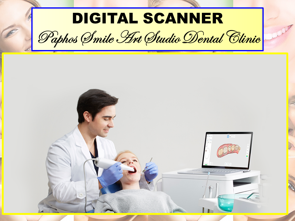
Digital intraoral scanners have become an ongoing trend in the dental industry and the popularity is only getting bigger. But what exactly is an intraoral scanner? Here we take a closer look at this incredible tool that makes all the difference, elevating the scanning experience for both doctors and patients to a whole new level.
What are intraoral scanners?
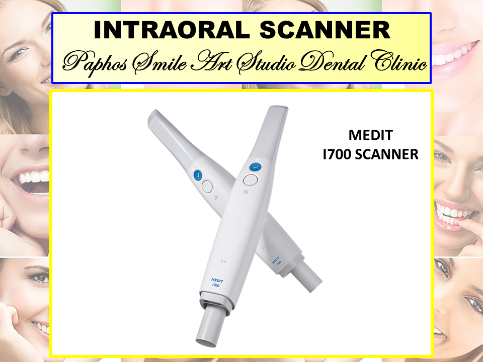
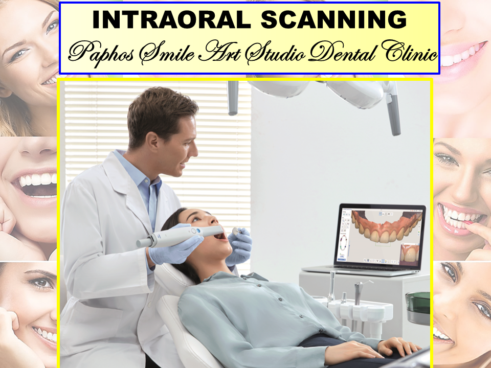
An intraoral scanner is a handheld device used to directly create digital impression data of the oral cavity. Light source from the scanner is projected onto the scan objects, such as full dental arches, and then a 3D model processed by the scanning software will be displayed in real-time on a touch screen. The device provides accurate details of the hard and soft tissues located in the oral area through high-quality images. It is becoming a more popular choice for clinics and dentists due to short lab turnaround times and excellent 3D image outputs.
Development of Intraoral scanners
In the 18th century, methods of taking impressions and making models were already available. At that time dentists developed many impression materials such as impregum, condensation /addition silicone, agar, alginate, etc. But impression making seems error-prone and is still uncomfortable to patients and time-consuming to dentists. To overcome these limitations, intraoral digital scanners have developed as an alternative to traditional impressions.
The advent of intraoral scanners has coincided with CAD/CAM technology development, bringing many benefits to the practitioners. In the 1970s, the concept of computer-aided design/ computer-aided manufacturing (CAD/CAM) was first introduced in dental applications by Dr. Francois Duret. By 1985, the first intraoral scanner became commercially available, used by labs to fabricate precise restorations. With the introduction of the first digital scanner, dentistry was offered an exciting alternative to conventional impressions. Although the scanners of the 80s are far from the modern versions we use today, digital technology has continued to evolve over the past decade, producing scanners that are faster, more accurate and smaller than ever before.
Today, intraoral scanners and CAD/CAM technology offer easier treatment planning, more intuitive workflow, simplified learning curves, improved case acceptance, produce more accurate results, and expand the types of treatments available. No wonder more and more dental practices are realizing the need to enter the digital world— the future of dentistry.
How do intraoral scanners work?

An intraoral scanner consists of a handheld camera wand, a computer, and software. The small, smooth wand is connected to a computer that runs custom software that processes the digital data sensed by the camera. The smaller the scanning wand, the more flexible it is in reaching deep into the oral area to capture accurate and precise data. The procedure is less likely to induce gag response, making the scanning experience more comfortable for patients.
In the beginning, dentists will insert the scanning wand into the patient's mouth and gently move it over the surface area of the teeth. The wand automatically captures the size and shape of each tooth. It only takes a minute or two to scan, and the system will be able to produce a detailed digital impression. (For example, Launca DL206 intraoral scanner takes less than 40 seconds to complete a full arch scan). The dentist can view the real-time images on the computer, which can be magnified and manipulated to enhance details. The data will be transmitted to labs to fabricate any needed appliances. With this instant feedback, the whole process will be more efficient, saving time and allowing dentists to diagnose more patients.
What are the advantages?
Enhanced patient scanning experience.
Digital scan reduces patient discomfort considerably because they do not have to endure the inconveniences and discomfort of traditional impressions, such as unpleasant impression trays and the possibility of gag reflex.
Time-saving and fast results
Reduces the chair time required for treatment and scan data can be sent immediately to the dental lab via the software. You can instantly connect with the dental Lab, reducing remakes and faster turnaround times compared to traditional practices.
Increased Accuracy
Intraoral scanners use the most advanced 3D imaging technologies that capture the exact shape and contours of the teeth. Enabling the dentist to have better scanning results and clearer teeth structure information of patients and give accurate and appropriate treatment.
Better patient education
It’s a more direct and transparent process. After a full-arch scan, dentists can use 3D imaging technology to detect and diagnose dental diseases by providing a magnified, high-resolution image and share it digitally with the patients on the screen. By seeing their oral condition almost instantly in the virtual world, patients will be able to communicate effectively with their doctors and more likely to move forward with treatment plans.

Let's Go Digital!
We believe you are aware that digital technology is an inevitable trend in all fields. It just brings so many benefits to both professionals and their clients, providing a simple, smooth and precise workflow that we all want. Professionals should keep up with the times and provide the best service to engage their clients. Choosing the right intraoral scanner is the first step towards digitalization in your practice, and it is crucial. Launca Medical has been dedicated to developing cost-effective, high-quality intraoral scanners.
Cancer screening tools
Fluorescence imaging can help dental professionals see abnormalities and signs of cancer that may not be visible to the naked eye. When diseases are diagnosed early with these tools, they can be treated at an earlier stage, giving the patient an improved prognosis and a shorter recovery time. A review published in the journal Oral Diseases confirms that this new tool can detect lesions and other potentially malignant disorders.
Digitally guided implant surgery
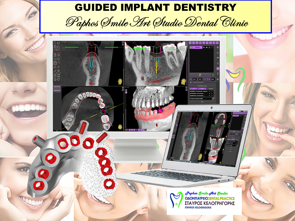
This relatively new innovation isn't widely used yet. Still, it helps dental professionals identify the most precise and effective way to place an implant in your specific jawbone structure using an intraoral scan. The American Academy of Implant Dentistry says that 3 million Americans have dental implants. That number is growing by 500,000 every year, so this innovation could ensure many people receive the best dental implant possible.
Learn more about dental implants and tooth bridges.
Information management
Not too long ago, dental records were all physical copies stored away in filing cabinets. A mix of technological innovation and federal regulation has motivated dental offices around the country to update their systems. This has improved scheduling, made it easier for your dental professional to access records right when they need them, improved workflows, and simplified patient information sharing between offices when necessary.
Digital technology in dentistry is accelerating rapidly, and dental health professionals who harness scientifically tested and proven advances in their field may be able to offer the best care. Digital dentistry is constantly providing dental professionals with ways in which they can help you in faster, safer, more comfortable, and more reliable ways than they ever have before. That's something you can smile about (IRL or digitally 😃).
Digital dentistry involves the use of dental technology or devices which use computer-based or digital components. Traditional dental mediums, on the other hand, use mechanical or electrical procedures.
Computer-based technology enables dental practices to further enhance patient care. Above all, digital dentistry eliminates manual steps of dental procedures. For this reason, dentists can provide an efficient and more automated treatment process.
Benefits of digital dentistry:
The development of new technologies and techniques in dentistry offer a variety of advantages for patients. This includes:
Accurate condition diagnoses
Dentists can use digital dentistry techniques to accurately identify, diagnose and treat oral diseases in its early stages. Unlike traditional x-ray images, digital radiography images and CBCT X-rays can be enlarged. For this reason, they can display an accurate representation of a patient’s teeth and oral anatomy.
Reduce time and costs
Instead of waiting days for a laboratory to create a restoration, a practitioner can have it ready on the same day. Therefore, the team can eliminate further appointments saving time and money. Furthermore, digital images are immediately available for labs and insurance providers. As well, patients do not have to wait for the images and can easily view them on the screen.
Enhanced patient experience
Digital imaging has also enabled our dentists to reduce the usage of traditional impression material which many patients find uncomfortable. Instead of taking impressions, your dentist can now easily scan your teeth with a 3D scanner. This means, you won’t have to bite into impression material or have trays inserted into your mouth. Overall, many patients find the use of the 3D scanner much more comfortable.
Improved communication
Digital imaging also allows our dentists to collaborate with other specialists and dental laboratories. This even can happen while the patient is still in the practice. Additionally, 3D teeth impressions can be sent off for instant feedback on procedures and planned restorations.
.jpg)